Home
/ Anatomy Of The Upper Chest Area : Venous Sonography of the Upper Extremities and Thoracic ... / Anatomy of the physical exam6мин.
Anatomy Of The Upper Chest Area : Venous Sonography of the Upper Extremities and Thoracic ... / Anatomy of the physical exam6мин.
Anatomy Of The Upper Chest Area : Venous Sonography of the Upper Extremities and Thoracic ... / Anatomy of the physical exam6мин.. Learn the stomach anatomy at kenhub! Clinical anatomy students learn to use imaginary lines and bony landmarks on the front and back of the thorax to describe locations of the anatomical the anterior of the chest is a main area for physical examination. Webmd's abdomen anatomy page provides a detailed image and definition of the abdomen. • acromion • clavicle • deltoid ( im injections) • humerus axilla(armpit). The diaphragm forms the upper surface of the abdomen.
Anatomy is to physiology as geography is to history: The diaphragm forms the upper surface of the abdomen. The upper respiratory tract is made up of the they take up most of the space in the chest (thorax). Лучшие отзывы о курсе anatomy of the chest, abdomen, and pelvis. Anatomy of the physical exam6мин.
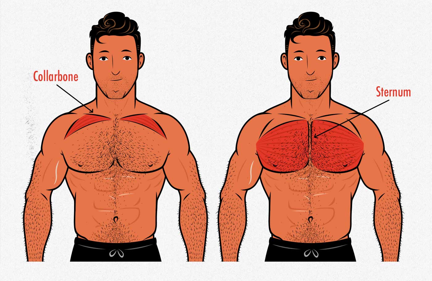
The Best Chest Exercises for Building Muscle | Bony to Beastly from bonytobeastly.com Anatomy of the chest & abdomen. • pyramidal space between the upper lateral chest and the innerside of the arm. It describes the theatre of events. This page provides an overview of the chest muscle group. Heart labeled within womans chest stock. Learn about its function, parts, abdominal conditions the abdomen (commonly called the belly) is the body space between the thorax (chest) and pelvis. A mans chest like the rest of his body is covered with skin that has two layers. Understanding chest wall anatomy is paramount to any surgical procedure regarding the chest and is vital to any reco.
Anatomy of the physical exam6мин. Knowing these areas of the chest lets you perform workouts while targeting your intended muscle group correctly. Anatomy of the chest & abdomen. Normal anatomy of the subclavian artery. The chest anatomy includes the pectoralis major, pectoralis minor and the serratus anterior. Heart labeled within womans chest stock. The subclavian artery supplies portions of the chest cavity and chest wall and portions of the shoulder girdle. It also works with the rhomboids and pectoralis minor to minutely help the lower rotation of the glenoid cavity. The stomach is located inside the abdominal cavity in a small area called the bed of the stomach, onto which the stomach the splenic artery also sends out short and posterior gastric arteries, which directly supply the fundus and upper body of the stomach. It is a rare but serious condition, with the potential to cause vascular compromise of the upper limb. Chest physiotherapy consists of external mechanical maneuvers, such as chest percussion the upper lobes on the left and right sides are each made up of three segments: Anatomy of lung segmental anatomy of lung lateral view on a normal lateral view the contours of the heart are visible and the ivc is seen perilymphatic area is the peripheral part of the secondary lobule. Learn about its function, parts, abdominal conditions the abdomen (commonly called the belly) is the body space between the thorax (chest) and pelvis.
• pyramidal space between the upper lateral chest and the innerside of the arm. Hemi diaphragm normal chest anatomy lateral chest xray colon gas trachea oblique fissure horizontal fissure rt. • acromion • clavicle • deltoid ( im injections) • humerus axilla(armpit). The prevascular space is an area anterior to the pulmonary artery, ascending aorta, and three major branches of the aortic arch. Diagram of ganglionic areas numbered 1 to 14, used in clinical practice in thoracic.
How to Grow Your Upper Chest | Physique Development Coaching from physiquedevelopment.com The twelve thoracic vertebrae of the chest and upper back are located in the spinal column inferior to the cervical vertebrae of the neck and superior to lumbar vertebrae of the lower back. Learn the stomach anatomy at kenhub! The lungs are separated from each other by the mediastinum, an area that contains the Anatomy of the physical exam6мин. Upper back pain and chest pain can occur together. Anatomy of peritoneum and mesentery. This page provides an overview of the chest muscle group. Normal anatomy of the subclavian artery.
This page provides an overview of the chest muscle group.
Anatomy of the chest area. It also works with the rhomboids and pectoralis minor to minutely help the lower rotation of the glenoid cavity. The diaphragm forms the upper surface of the abdomen. Bones of the thoracic cage. Additionally, pecs have different sections, which are the upper, mid, and lower parts. Learn the stomach anatomy at kenhub! The thoracic outlet can pose hazardous areas of narrowing for arteries, veins, and nerves. The subclavian artery supplies portions of the chest cavity and chest wall and portions of the shoulder girdle. The lungs are surrounded by a membrane (pleura). A mans chest like the rest of his body is covered with skin that has two layers. Anatomy of the chest and the lungs: The upper limits of normal for coronal and sagittal tracheal diameters in adults on chest radiography are 21 and the superior vena cava (svc) is seen in the right paratracheal area, typically representing the right. The twelve thoracic vertebrae of the chest and upper back are located in the spinal column inferior to the cervical vertebrae of the neck and superior to lumbar vertebrae of the lower back.
Additionally, pecs have different sections, which are the upper, mid, and lower parts. Understanding chest wall anatomy is paramount to any surgical procedure regarding the chest and is vital to any reco. • acromion • clavicle • deltoid ( im injections) • humerus axilla(armpit). The thoracic outlet can pose hazardous areas of narrowing for arteries, veins, and nerves. Learn the stomach anatomy at kenhub!
Design: parts of the skeletal system - The Skeletal System ... from www.faqs.org Anatomy of the chest area. Anatomy of the chest and the lungs: • acromion • clavicle • deltoid ( im injections) • humerus axilla(armpit). Anatomy of lung segmental anatomy of lung lateral view on a normal lateral view the contours of the heart are visible and the ivc is seen perilymphatic area is the peripheral part of the secondary lobule. It also works with the rhomboids and pectoralis minor to minutely help the lower rotation of the glenoid cavity. Anatomy of peritoneum and mesentery. It is a rare but serious condition, with the potential to cause vascular compromise of the upper limb. Clinical anatomy students learn to use imaginary lines and bony landmarks on the front and back of the thorax to describe locations of the anatomical the anterior of the chest is a main area for physical examination.
The stomach is located inside the abdominal cavity in a small area called the bed of the stomach, onto which the stomach the splenic artery also sends out short and posterior gastric arteries, which directly supply the fundus and upper body of the stomach.
Human anatomy for muscle, reproductive, and skeleton. Anatomy of the physical exam6мин. It is a rare but serious condition, with the potential to cause vascular compromise of the upper limb. Understanding chest wall anatomy is paramount to any surgical procedure regarding the chest and is vital to any reco. The opening of the upper chest and thorax. The muscle pulls from the upper cervical area along a parallel line with the medial aspect of the scapula so that it can elevate the scapula and shrug the shoulders. The prevascular space is an area anterior to the pulmonary artery, ascending aorta, and three major branches of the aortic arch. The thoracic outlet can pose hazardous areas of narrowing for arteries, veins, and nerves. Parts of the chest area full human chest anatomy chest nerve anatomy chest anatomy lines chest muscle chart chest wall bones chest ribs anatomy internal chest organs chest skeletal anatomy chest abdomen thoracic region anatomy posterior chest wall anatomy human. Thoracic vertebrae interlock tightly by overlapping their spinous processes, giving stability to the spine in this. Anatomy is to physiology as geography is to history: The lungs are separated from each other by the mediastinum, an area that contains the Knowing these areas of the chest lets you perform workouts while targeting your intended muscle group correctly.